 Oded Bahat Research by Dr. Oded Bahat
Related Articles
Growth is something that occurs from infancy to 18 years of age. After that, it was thought, that growth ceases. Dentists used this fact to establish placement for dental implants. With set locations in mind, it was possible to establish a fixed occlusion and to ensure that patients were comfortable esthetically and functionally with the implants. However, it has since been established that growth continues all through life. In fact, the maxilla and mandible continue growing even after aging is complete. In fact, teeth positioning and occlusion also changes all through life. This premise means that dentists have to foretell and compensate for this change while planning implant placement in partially or completely edentulous patients. In this article, we will take a closer look at potential age-related changes and how to adjust for these changes.
Common Changes Associated With Aging And How To Deal With Them
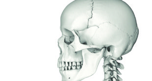
The most obvious visible changes in the craniofacial region occur due to constant use, aging, and gravity but aging changes are also seen at the microscopic level. Facial bone density decreases, teeth migrate mesially, the face grows longer, the mandibular angle increases, ramus height and mandibular height decrease, and facial height increases with aging. The exact amount of change varies according to gender but men show a downward growth in the posterior region while women show an increase in the mandibular angle. Both men and women demonstrate mesial movement of teeth and a decrease in arch circumference and arch length.
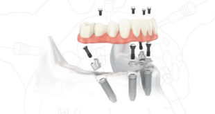
As a result, 3-D bone morphology imaging may not be enough to assess bone density for implant placement. It does become necessary to carry out bone augmentation procedures to build a foundation for the implants. At present, six different kinds of augmentation procedures are carried out by surgeons to acquire bony support: onlay block grafts, guided bone regeneration, sinus floor elevation, distraction osteogenesis, interpositional grafts and alveolar ridge expansion. However, the most common technique preferred involves the use of the patient’s own bone with a tissue matrix or graft. This kind of in-situ tissue regeneration is perfect as it allows for a significant amount of bone development. Implant sites have a reduced risk of perforation or exfoliation and there is a significantly lower risk as compared to open autogenous bone harvest transplants.
Conclusion
It is not possible to predict the exact type, variety and extent of craniofacial changes that will occur in a particular individual. It will become necessary for the dental surgeon to carry out a risk assessment and a complete dental evaluation of the patient before treatment. The patient should be informed that esthetics may worsen over time, particularly in patients who received their implants while young. It may become necessary to restore or replace implants after some time due to alveolar changes and tooth drifting. Researchers agree that long term research studies will be required to assess the potential of grafting and age-related changes on edentulous patients.
The information on this post is meant for informational purposes only, and is not to be construed as medical advice. If you are experiencing a dental emergency, please contact a doctor immediately.